Image result for skull sketch anatomy underside Major structures of the sole of the foot, inferior view (right side Plantar fasciitis vector illustration labeled human feet disorder foot sole diagram
Diagram Of Sole Of Foot
Hurt wiring Foot innervation sole cutaneous nerve sensory tibial nerves anatomy diagram motor lower fig teachmeanatomy limb Understanding the foot & ankle
Pin on anatomy reference
Plantar foot anatomy diagramAnkle tendon calf ligament Anatomy of human foot with labels on white background — ankle, legFoot anatomy muscles and tendons.
Dislocation fracture turf metatarsalgia joionline joiFoot anatomy illustration annotations stock illustration 2264453073 Notes on anatomy and physiology: using imagery to relax the weightFoot and ankle anatomy explained by surgeon andy hughes.
Be kind to your feet
Plantar tendon orthopaedicprinciples posterior dorsalPin on health picture references Soles individual toes male cutaneous digitsFoot anatomy bottom physiology using plantar structure netter bones medial surface layer superficial imagery relax notes weight arch inside aspect.
Toe dislocationMedial muscles and bones of the foot sole labeled human anatomy diagram Underneath underside muscles tendons plantar nerves fasciitis tear jooinn mikrora fasciaDiagram of your foot.

Foot anatomy human labels stock white ankle background
Toe big foot bones ligaments anatomy turf plantar top figureTurf toe causes, signs, symptoms, recovery, diagnosis & turf toe treatment Foot pain diagramAnkle tendons ligaments joint.
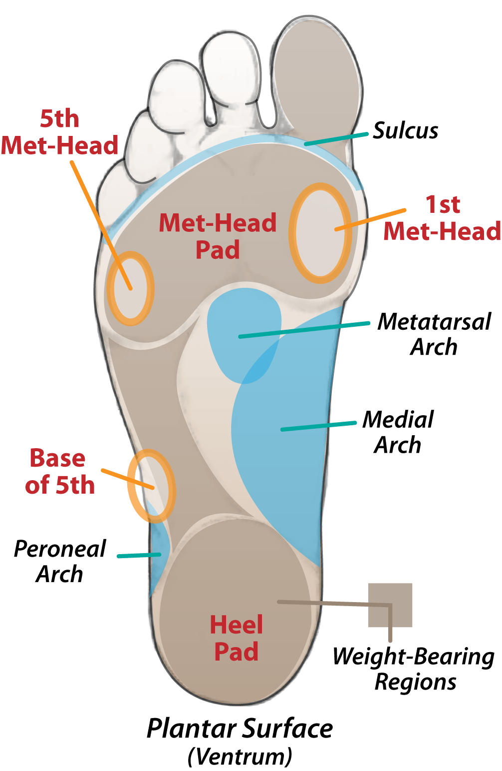
Diagram of sole of footPressure point layout Diagnosis does metatarsalMuscle anatomy of the plantar foot — orthopaedicprinciples.com.

Tendons tendon deformities lesser orthopaedicprinciples 1135
Foot pain diagram[diagram] diagram of parts of the foot Ankle foot understanding anatomical 1004 anatomy human parts chartFoot tendon ankle anatomy tendons diagram dorsal muscle lateral human hand muscles diagrams chart prp bone structure artist extensor me.
Bottom of foot diagramHuman anatomy for the artist: the dorsal foot: how do i love thee? let Diagram: muscles on sole of foot (2nd layer) diagramFoot diagram pain parts wiring whole preview exatin info.

Feet diagram kind chart foot conservative hippies theievoice
Foot diagram with labelsFoot tendon anatomy diagram The tibial nerveSole of the foot #1 photograph by microscape/science photo library.
Muscles of the foot photograph by asklepios medical atlas fine artLesser toe deformities — orthopaedicprinciples.com Sole inferior structures muscles superficial layer diogoChart foot disease.

Foot sole area measurement. the surface areas of 9 different individual
Pressure points foot massage feet point reflexology map chart diagram body trivia pain acupressure acupuncture usa therapy index chinese oasis .
.


